Peripheral and central parts of the speech apparatus
Chapter 3. ANATOMY, PHYSIOLOGY AND PATHOLOGY OF VOICE AND SPEECH FORMATION
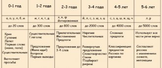
Speech is one of the basic functions of a person, necessary for him to lead a full life. The natural period of speech formation is the first three years of a baby’s life, and if at this time favorable conditions for the formation of this mental function are not created, in the future it will be much more difficult, and sometimes even impossible, to compensate for it!
The development of children's speech and vocabulary, mastery of the riches of their native language is one of the main elements of personality formation, the development of the developed values of national culture, is closely related to mental, moral, aesthetic development and is a priority in the language education and training of children, especially preschoolers.
Research by many scientists in this area suggests that for the development of a child’s speech, normal maturation and functioning of the central nervous system, the normal state of higher mental processes (memory, attention, thinking, imagination), as well as the physical condition of the child are necessary. However, even with all the necessary prerequisites, a child will never speak without a speech environment.
On the other hand, the formation of higher mental functions and feelings (moral, aesthetic, intellectual) in humans is carried out only through speech. If a person’s speech does not develop (children are “Mowgli”), then neither moral, nor aesthetic, nor intellectual feelings, even in the most primitive form, are formed. The development of perception, thinking, and memory also suffers significantly, because the basis of the second signaling system, which plays a leading role in the development of these higher mental functions, is the word, oral and written speech, as a system of meaningful speech signals characteristic only of humans.
The concept of language does not coincide with the concept of speech activity in general, since language is only a certain part, admittedly the most important part, of speech activity. Speech activity is a product of the functioning of the speech organs, phylogenetically developed and inherent in any Homo sapiens (reasonable person). All people tend to reproduce the same sounds, but each society forms speech (verbal) signs from them, denoting certain concepts, which become the language of communication of the limited population in which they have developed, because they are perceived by everyone in the same semantic sense. meaning. There is a parallel formation of speech, as the ability to independently pronounce the necessary sounds and transform them into words, and language, as a means of communication between people. A language is common to the people who speak it, but speech is always individual and unique, and even over the telephone, when the frequency range of the transmitted speech signal is significantly limited, we recognize familiar people.
Thus, if sound pronunciation (unlike, for example, fish) is an innate biological component of humans and many animals, then sound creation (i.e. learning to consciously pronounce the entire range of necessary sounds), which forms the basis of colloquial speech, is its phylogenetically acquired biological component, and language is a social product of speech ability. Language is a treasure deposited by the practice of speech in all who belong to the same community (society). As an ingredient of speech, it permeates all speech and all its aspects.
If the development of speech is associated with neuro-motor stimulation of the speech-motor apparatus, then the development of language is with an increase in vocabulary, the formation of the correct grammatical structure of speech, the ability to adequately use vocabulary in thinking (inner speech) and communication with others (external speech). In this regard, emerging problems of speech development are mainly problems of doctors and speech therapists, and the development of language as the basis of speech communication is a purely pedagogical task.
The anatomical structure and physical characteristics of the human articulatory organs are well adapted to the production of human speech. And, perhaps, vice versa - human speech in the form in which it was formed in the process of evolution is determined by the physical characteristics of the human organs of articulation and the limitations that are associated with the possibilities of their change and movement in space and time.
Physiologically, speech is a complex motor act carried out according to the mechanism of conditioned reflex activity. It is formed on the basis of kinesthetic stimuli emanating from the speech muscles, including the muscles of the larynx and respiratory muscles.
The sound expressiveness of speech is controlled using an auditory analyzer, the normal activity of which plays a very important role in the development of speech in a child. Speech acquisition occurs in the process of interaction of the child with the environment, in particular with the speech environment, which is a source of imitation for the child. In this case, the child uses not only a sound, but also a visual analyzer, imitating the corresponding movements of the lips, tongue, etc. The kinesthetic stimuli that arise in this case enter the corresponding area of the cerebral cortex. A conditioned reflex connection is established and consolidated between three analyzers (motor, auditory and visual), ensuring the further development of normal speech activity.
Observations on the development of speech in blind children show that the role of the visual analyzer in the formation of speech is secondary, since speech in such children, although it has some features, develops, in general, normally and, as a rule, without special outside intervention. Thus, the development of speech is associated mainly with the activity of the auditory and motor analyzers.
Speech reflexes are associated with the activity of various parts of the brain. Therefore, in the speech apparatus there are two closely interconnected parts: the central (regulatory) and peripheral (executive) speech apparatus (Fig. 10).
Rice. 10. Structure of the speech apparatus
The central speech apparatus includes:
- the cortical ends of the analyzers (primarily auditory, visual and motor) involved in the speech act. The cortical end of the auditory analyzer is located in both temporal lobes, the visual one is in the occipital lobes, and the cortical part of the motor analyzer, which ensures the functioning of the muscles of the jaws, lips, tongue, soft palate, larynx, which also takes part in the speech act, is located in the lower parts of these convolutions;
- The sensory speech motor apparatus is represented by proprioceptors located inside the muscles and tendons involved in the speech act and excited by contractions of the speech muscles. Baroreceptors are located in the pharynx and are excited by changes in pressure on them when pronouncing speech sounds;
- afferent (centripetal) pathways begin in proprioceptors and baroreceptors, and carry the information received from them to the cerebral cortex. The centripetal path plays the role of a general regulator of all activities of the speech organs;
- cortical speech centers are located in the frontal, temporal, parietal and occipital lobes of the predominantly left hemisphere of the brain. The emotional-figurative component of speech depends on the participation of the right hemisphere.
The frontal gyri (inferior) are the motor area and are involved in the formation of one's own oral speech. The temporal gyri (superior) are the speech-auditory area where sound stimuli are received. Thanks to this, the process of perceiving someone else’s speech is carried out. The parietal lobe of the cerebral cortex is important for understanding speech. The occipital lobe is a visual area and ensures the assimilation of written speech (perception of letter images when reading and writing) and articulation in adults, which also plays an important role in the development of a child’s speech; specific speech centers (sensory - Wernicke and motor - Broca), responsible for fine sensory analysis and neuromuscular coordination of speech (Fig. 11)
Wernicke's auditory sensory (sensitive) speech center is located in the posterior part of the left superior temporal gyrus. When it is damaged or diseased, disturbances in sound perception occur. Sensory aphasia occurs, in which it becomes impossible to distinguish speech elements (phonemes and words) by ear, and, consequently, to understand speech, although hearing acuity and the ability to distinguish non-speech sounds remain normal.
Broca's auditory motor speech center is located in the posterior part of the second and third frontal gyri of the left hemisphere. Damage or disease of the motor center of speech leads to disruption of the analysis and synthesis of kinesthetic (motor) stimuli that occur when pronouncing speech sounds. Motor aphasia occurs, in which it becomes impossible to pronounce words and phrases, although movements of the speech organs not related to speech activity (movements of the tongue and lips, opening and closing the mouth, chewing, swallowing, etc.) are not impaired.
Rice. 11. Areas of motor and auditory analyzers
speech in the cerebral cortex
1 – motor analyzer (anterocentral gyrus;
2 – motor (motor) speech center (Broca);
3 – sensory speech center (Wernicke)
§ subcortical nodes and nuclei of the brainstem (primarily the medulla oblongata), control the rhythm, tempo and expressiveness of speech;
§ efferent (centrifugal) pathways connect the cerebral cortex with the respiratory, vocal and articulatory muscles that provide the speech act. They begin in the cerebral cortex in Broca's center.
The efferent pathways also include cranial nerves, which originate in the nuclei of the brain stem and innervate all organs of the peripheral speech apparatus.
The trigeminal nerve innervates the muscles that move the lower jaw; facial nerve - facial muscles, including muscles that carry out lip movements, puffing and retraction of the cheeks; glossopharyngeal and vagus nerves - muscles of the larynx and vocal folds, pharynx and soft palate. In addition, the glossopharyngeal nerve is the sensory nerve of the tongue, and the vagus nerve innervates the muscles of the respiratory and cardiac organs. The accessory nerve innervates the muscles of the neck, and the hypoglossal nerve supplies the muscles of the tongue with motor nerves and gives it the possibility of a variety of movements.
The peripheral speech apparatus consists of three sections: 1) respiratory; 2) voice; 3) articulatory (or sound-reproducing).
on the. This is the supplier of air for sound formation, since speech sounds from a physical point of view are nothing more than mechanical vibrations of exhaled air of various frequencies and strengths that arise in the subsequent peripheral part of the speech apparatus - the vocal apparatus. The respiratory section includes the chest with the lungs, bronchi and trachea (Fig. 12). The role of the respiratory section in human speech production is one to one reminiscent of the role of the bellows of a wind musical instrument - org
1 – nasal cavity; 2 – oral cavity; 3 – palate; 4 – nasopharynx; 6 – oral part of the pharynx; 6 – epiglottis; 7 – hyoid bone; 8 – larynx; 9 – esophagus; 10 – trachea; 11 – apex of the left lung; 12 – left lung; 13 – left bronchus; 14-15 – pulmonary vesicles (alveoli); 16 – right bronchus; 17 – right lung
Rice. 12. Airways. Showing the branches of the bronchi in the lungs (bronchial tree)
The vocal section consists of the larynx with the vocal folds located in it (Fig. 13–14). The larynx is a wide, short tube consisting of cartilage and soft tissue. It is located in the front of the neck and can be felt through the skin from the front and sides, especially in thin people.
From above, the larynx passes into the pharynx, from below - into the windpipe (trachea) - (Fig. 10). Two pathways cross in the pharynx - the respiratory and the digestive. The role of the “arrows” in this crossing is played by the soft palate and epiglottis (Fig. 15).
Rice. 13. Cartilaginous skeleton of the larynx: (A – front; B – back) 1 – trachea; 2 – cricoid cartilage; 3 – thyroid cartilage; 4 – arytenoid cartilages; 5 – epiglottis
Rice. 14. Vertical section through the larynx (the anterior half of the larynx is visible from the inside)
1 – epiglottis; 2 – aryepiglottic fold; 3 – thyroid cartilage; 4 – false vocal cord; 5 – blinking ventricle; 6 – true vocal cord (fold); 7 – cricoid cartilage; 8 – trachea
The soft palate serves as a posterior continuation of the hard palate; it is a muscular formation covered with a mucous membrane. The back part of the soft palate is called the velum palatine. When the palatine muscles relax, the palatine curtain hangs down freely, and when they contract (which is observed during the act of swallowing), it rises upward and backward, blocking the entrance to the nasopharynx. In the middle of the velum palatine there is an elongated process - the uvula.
The epiglottis consists of cartilaginous tissue shaped like a tongue or petal. Its front surface faces the tongue, and its back surface faces the larynx. The epiglottis serves as a valve: descending during the swallowing movement, it closes the entrance to the larynx and protects its cavity from the entry of food and saliva (Fig. 15).
Men have a larger larynx and longer and thicker vocal folds than women. The vocal folds with their mass almost completely cover the lumen of the larynx, leaving a relatively narrow glottis.
In children, the larynx is small and grows unevenly at different periods. Its noticeable growth occurs at the age of 5-7 years, and then during puberty: in girls at 12-13 years old, in boys at 13-15 years old. At this time, the size of the larynx in girls smoothly increases by one third, and in boys this process is “explosive” in nature: the Adam’s apple begins to quickly appear, and the significantly (2/3) increased vocal folds lead to a “change of voice” - a change in its timbre .
Rice. 15. Position of the soft palate and epiglottis during breathing (A) and swallowing (B)
1 – soft palate; 2 – epiglottis; 3 – trachea; 4 – esophagus
The activity of the vocal cords is associated with the paired internal muscles of the larynx (Fig. 16):
The muscles that stretch the vocal cords are the thyroarytenoid (or vocal) and cricothyroid muscles. The former, together with the mucous membrane covering them, form the true vocal cords (folds), between which there is a glottis. When the thyroarytenoid muscle contracts, the vocal cords stretch and, increasing in diameter, somewhat narrow the glottis. When the cricothyroid muscle contracts, due to the inclination of the thyroid cartilage, tension on the vocal cords also occurs;
Rice. 16. Muscles of the larynx
A – posterior: 1 – posterior cricoarytenoid muscle; 2 – transverse interarytenoid muscle; 3 – oblique interarytenoid muscles
B – lateral view: 1 – posterior cricoarytenoid muscle; 2 – lateral cricoarytenoid muscle; 3 – thyroarytenoid muscle
Rice. 17. Scheme of action of the posterior cricoarytenoid muscle
1 – true vocal fold; 2 – vocal process; 3 – muscle process
- The group of muscles that expand the glottis includes only one muscle - the posterior crico-arytenoid, called for short simply the posterior muscle of the larynx. During its contraction, it rotates the arytenoid cartilages around a vertical axis, as a result of which the vocal processes of these cartilages, together with the posterior ends of the true vocal cords attached to them, diverge to the sides and open the glottis (Fig. 17);
- The group of muscles that narrow the glottis includes: the lateral cricoarytenoid muscle, which serves as an antagonist of the posterior muscle, and the transverse arytenoid, or simply the transverse muscle, which is the only unpaired muscle of the larynx. During its contraction, it brings the arytenoid cartilages closer together, thereby contributing to the closure of the glottis. The action of this muscle is complemented by the right and left oblique arytenoid muscles, which intersect with each other.
The larynx is innervated by the sensory and motor branches of the vagus nerve.
Articulation department. The main organs of articulation are the tongue, lips, jaws (upper and lower), hard and soft palates, and alveoli. Of these, the tongue, lips, soft palate and lower jaw are mobile, the rest are fixed (Fig. 18).
The main organ of articulation is the tongue. The tongue is a massive muscular organ. When the jaws are closed, it fills almost the entire oral cavity. The front part of the tongue is mobile, the back part is fixed and is called the root of the tongue. The movable part of the tongue is divided into the tip, the leading edge (blade), the lateral edges and the back. The complexly intertwined system of tongue muscles (Fig. 19), the variety of their attachment points, provide the ability to change the shape, position and degree of tension of the tongue within a wide range. This not only plays a big role in the process of pronunciation of speech sounds, since the tongue is involved in the formation of all vowels and almost all consonants (except labials), but also ensures the processes of chewing and swallowing.
Rice. 18. Schematic profile of the organs of articulation
1 – lips; 2 – incisors; 3 – alveoli; 4 – hard palate; 5 – soft palate; 6 – vocal folds; 7 – root of the tongue; 8 – back of the tongue; 9 – tip of tongue
Rice. 19. Muscles of the tongue
1 – longitudinal muscle of the tongue; 2 – genioglossus muscle; 3 – hyoid bone; 4 – hypoglossus muscle; 5 – styloglossus muscle; 6 – styloid process
The muscles of the tongue (Fig. 19) are divided into two groups. The muscles of one group begin from the bony skeleton and end in one place or another on the inner surface of the mucous membrane of the tongue. This group of muscles ensures the movement of the tongue as a whole. The muscles of the other group are attached at both ends to various parts of the mucous membrane and, when contracted, change the shape and position of individual parts of the tongue. All muscles of the tongue are paired.
The first group of muscles of the tongue includes:
1. genioglossus muscle – pushes the tongue forward (sticking the tongue out of the mouth);
2. sublingual-lingual – pushes the tongue downwards;
3. styloglossus muscle - being an antagonist of the first (genioglossus), it retracts the tongue into the oral cavity.
The second group of muscles of the tongue includes:
1. superior longitudinal muscle - when contracted, it shortens the tongue and bends its tip upward;
2. inferior longitudinal muscle - contracting, hunches the tongue and bends its tip downwards;
3. transverse muscle of the tongue – reduces the transverse size of the tongue (narrows it and sharpens it).
The tongue receives motor innervation from the hypoglossal nerve (XII pair of cranial nerves), sensory innervation from the trigeminal nerve, and gustatory innervation from the glossopharyngeal nerve (IX pair).
The mucous membrane of the lower surface of the tongue, passing to the bottom of the oral cavity, forms a fold along the midline - the so-called frenulum of the tongue. In some cases, the frenulum, being insufficiently elastic or too short, limits the movement of the tongue, making it difficult to articulate.
An important role in the formation of speech sounds also belongs to the lower jaw, lips, teeth, hard and soft palate, and alveoli. Articulation consists in the fact that the listed organs form slits, or closures, that occur when the tongue approaches or touches the palate, alveoli, teeth, as well as when the lips are compressed or pressed against the teeth.
The formation of speech sounds also largely depends on the articulation of the lips, provided by part of the facial muscle apparatus (Fig. 20).
In addition to the orbicularis oris muscle, which is located in the thickness of the lips and, when contracted, presses the lips together, there are numerous muscles around the oral opening that provide various movements of the lips: the levator labii superioris muscle, the zygomatic minor muscle, the zygomaticus major muscle, the Santorini muscle of laughter and etc. The system of muscles that change the shape of the oral opening should also include the group of masticatory muscles. For example, the masseter and temporal muscles lift the lowered lower jaw; The pterygoid muscles, contracting simultaneously on both sides, push the jaw forward, and when they contract on one side, the jaw moves in the opposite direction. The lowering of the lower jaw when opening the mouth occurs mainly due to its own gravity (the chewing muscles are relaxed) and, partly, due to contraction of the neck muscles.
1–2 – muscles that lift the upper lip and wing of the nose; 3 – zygomatic minor muscle; 4 – muscle that lifts the anguli oris; 5 – zygomaticus major muscle; 6 – buccal muscle (trumpet muscle); 7 – orbicularis oris muscle; 8 – Santorini muscle of laughter; 9 – muscle that lowers the lower lip; 10 – muscle that lowers the angle of the mouth; 11 – chewing muscle
Rice. 20. Muscles of lips and cheeks
The muscles of the lips and cheeks are innervated by the facial nerve, and the muscles of mastication receive innervation from the motor root of the trigeminal nerve.
New ears
Hearing is the most complex tool for understanding the world. If for some reason nature did not set it up (we are talking primarily about genetic defects) or it was damaged as a result of injury or illness, humanity has learned to correct the deficiency by installing cochlear implants. But it is not immediately possible to bring the perception of sounds to the maximum possible with them.
Unlike hearing aids, which essentially physically amplify sound, cochlear implants involve electrical stimulation of the auditory nerve. The process of setting up a speech processor, even to an amateur’s eye, differs from a simple “turn the wheel and you hear everything.” This is a multi-stage, responsible (especially if we are talking about a baby who cannot talk about his feelings) and a very difficult task.
A cochlear implant, simply put, consists of:
- The internal part – the implant itself. It is assumed that it is implanted into a person once in a lifetime and forever (one implant replaces one ear).
- External – speech processor. This is a high-tech electronic device that picks up sounds, converts them into electrical signals and sends them to the internal structures of the implant. From there, sounds already distributed into high, medium and low frequencies enter the auditory nerve and go to the cerebral cortex. Speech processors can and should be changed over time.
The setting consists of the fact that the specialist, with feedback from the patient, determines the thresholds for electrical stimulation of the remaining auditory nerve fibers and sets the most effective and comfortable parameters for the person.
The result that everyone strives for is grade I hearing loss or a healthy standard of sound perception. In medical terms, the thresholds for the perception of tones across the entire scale during audiometry in a free sound field should be within the range of 25 to 40 dB.
Let's take a closer look.


